Bandung, June 2025 — A significant milestone has been reached in ophthalmic imaging through a dissertation-based study recently published in IEEE Access (DOI: 10.1109/ACCESS.2025.3573213). The paper, titled “Depth Map Reconstruction for Scleral Bleb Models Using Structured Light in Light Field Imaging”, presents an innovative method for three-dimensional depth map reconstruction targeting scleral bleb surfaces—an essential component in evaluating the success of trabeculectomy, a surgical procedure commonly used to treat glaucoma.
The study is part of an ongoing doctoral research project under STEI at Institut Teknologi Bandung (ITB). One of the supervisors and co-author of this research is Prof. Andriyan Bayu Suksmono, principal researcher at PPTIK ITB.
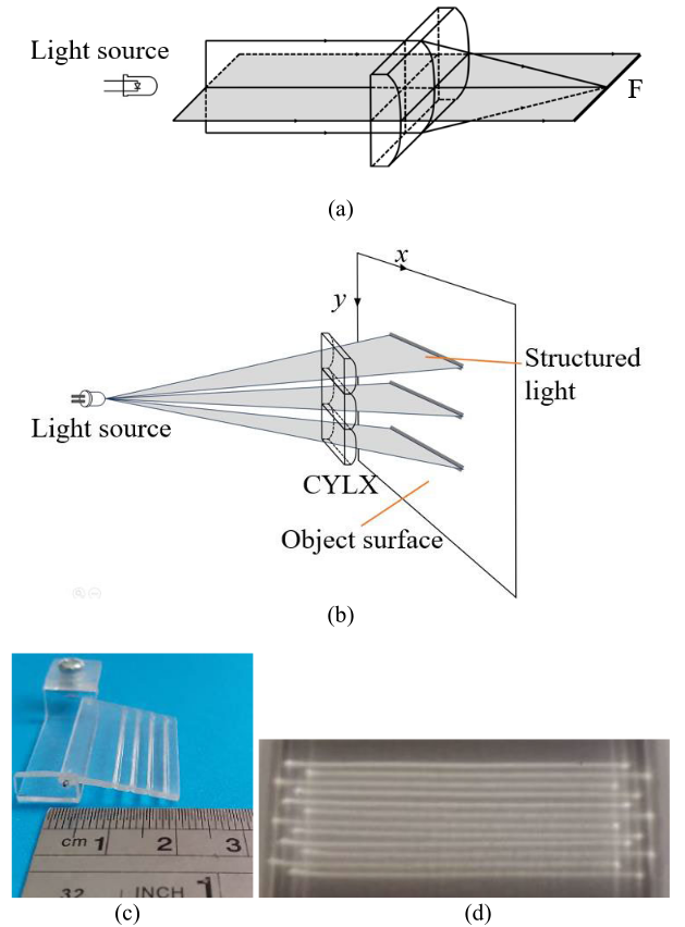
Figure 1. (a) Light beam from 1 CYLX, (b) light beam from three parallel CYLX, (c) the fabricated CYLX, and (d) generated structured light.
Solving Depth Reconstruction Challenges in Eye Imaging
Post-trabeculectomy blebs are notoriously difficult to analyze due to their white, glossy, and textureless surfaces, which cause conventional depth reconstruction methods to fail. This research introduces a structured light-assisted light field imaging system, in which a consumer-grade light field camera is integrated with a standard slit lamp. The structured light, generated via custom-fabricated cylindrical lenses, enhances contour visibility on the bleb surface, significantly improving depth accuracy.
Algorithmic Innovation with Clinical Relevance
The proposed method combines two core innovations:
– A reliability-guided disparity propagation (RGDP) method to refine initial depth estimations.
– A second-stage optimization using Markov Random Field (MRF), integrating color segmentation and edge detection from the central sub-aperture image.
The result is a high-fidelity, edge-preserving depth map reconstruction even under low-texture and variable lighting conditions.
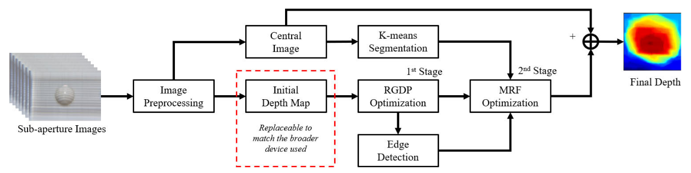
Figure 2. The proposed method diagram: Sub-aperture images preprocessing, followed by depth map generation via the defocus method. The depth map is optimized in two stages: first, using RGDP [34], and second, using MRF with segmentation and edge detection. The final depth map is produced by fusing the optimized result with the central sub-aperture image.
Tested on Real Eye Models and Public Datasets
This method was validated using both:
– Synthetic datasets from the Heidelberg Collaboratory for Image Processing (HCI), and
– Real-world experiments using a physical eye phantom designed to mimic human ocular anatomy.
Across all scenarios, the method outperformed existing techniques in terms of SSIM (Structural Similarity Index), SNR (Signal-to-Noise Ratio), and visual realism.
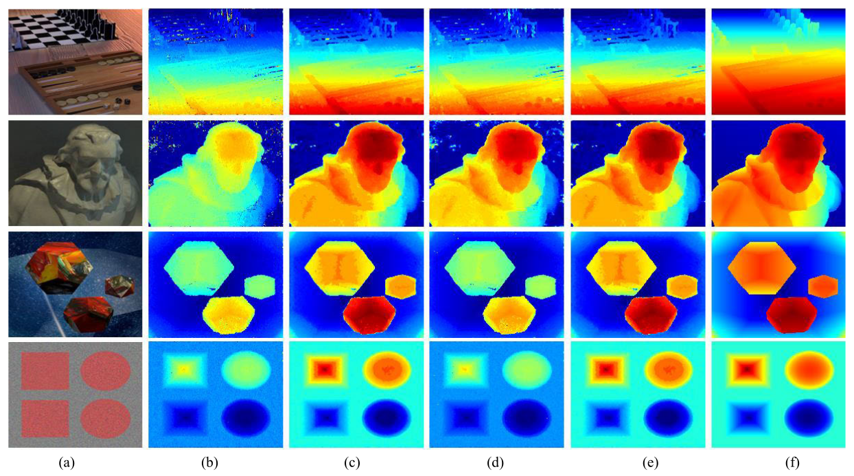
Figure 3. Experiment results on the dataset. Column (a) from top to bottom shows the sub-aperture central images of the boardgames, platonic, cotton, and pyramids, respectively. The subsequent columns illustrate the reconstructed depth map using various methods: (b) defocus method, (c) MRF-only optimization, (d) the first optimization, RGDP, (e) the proposed method, and (f) ground truth.
Towards Practical Ophthalmic Applications
Though not yet real-time, the system’s end-to-end processing time (approximately 4–5 minutes) is deemed acceptable for clinical settings. The proposed technique may soon support automatic grading scales such as IBAGS, offering 3D measurements for bleb height and extent—parameters traditionally assessed subjectively via 2D observation.
By leveraging accessible imaging tools and efficient computational methods, this dissertation research contributes directly to advancing intelligent, real-world solutions for healthcare.